http://www.ncbi.nlm.nih.gov/pubmed/25934284
Abstract
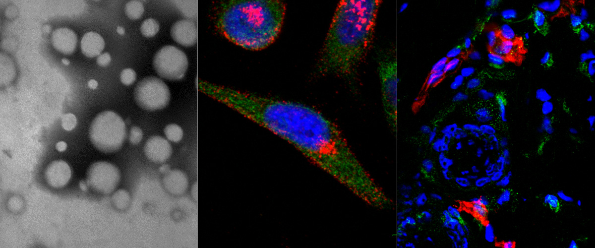
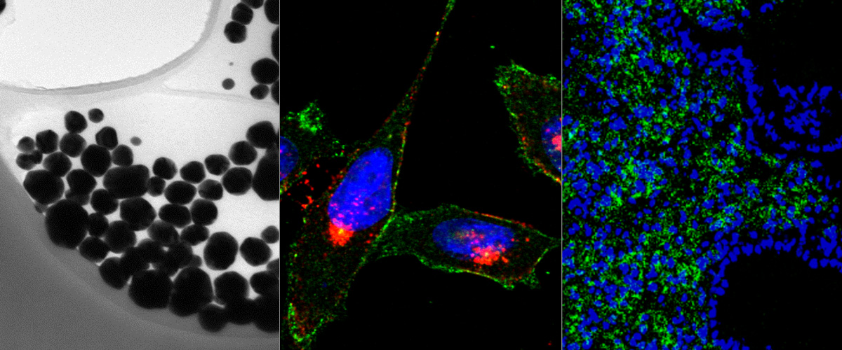
Ultrasound molecular imaging has great potential to impact early disease diagnosis, evaluation of disease progression and the development of target-specific therapy. In this paper, two neuropilin-1 (NRP) targeted peptides, CRPPR and ATWLPPR, were conjugated onto the surface of lipid microbubbles (MBs) to evaluate molecular imaging of tumor angiogenesis in a breast cancer model. Development of a molecular imaging agent using CRPPR has particular importance due to the previously demonstrated internalizing capability of this and similar ligands. In vitro, CRPPR MBs bound to an NRP-expressing cell line 2.6 and 15.6 times more than ATWLPPR MBs and non-targeted (NT) MBs, respectively, and the binding was inhibited by pretreating the cells with an NRP antibody. In vivo, the backscattered intensity within the tumor, relative to nearby vasculature, increased over time during the ∼6 min circulation of the CRPPR-targeted contrast agents providing high contrast images of angiogenic tumors. Approximately 67% of the initial signal from CRPPR MBs remained bound after the majority of circulating MBs had cleared (8 min), 8 and 4.5 times greater than ATWLPPR and NT MBs, respectively. Finally, at 7-21 days after the first injection, we found that CRPPR MBs cleared faster from circulation and tumor accumulation was reduced likely due to a complement-mediated recognition of the targeted microbubble and a decrease in angiogenic vasculature, respectively. In summary, we find that CRPPR MBs specifically bind to NRP-expressing cells and provide an effective new agent for molecular imaging of angiogenesis.