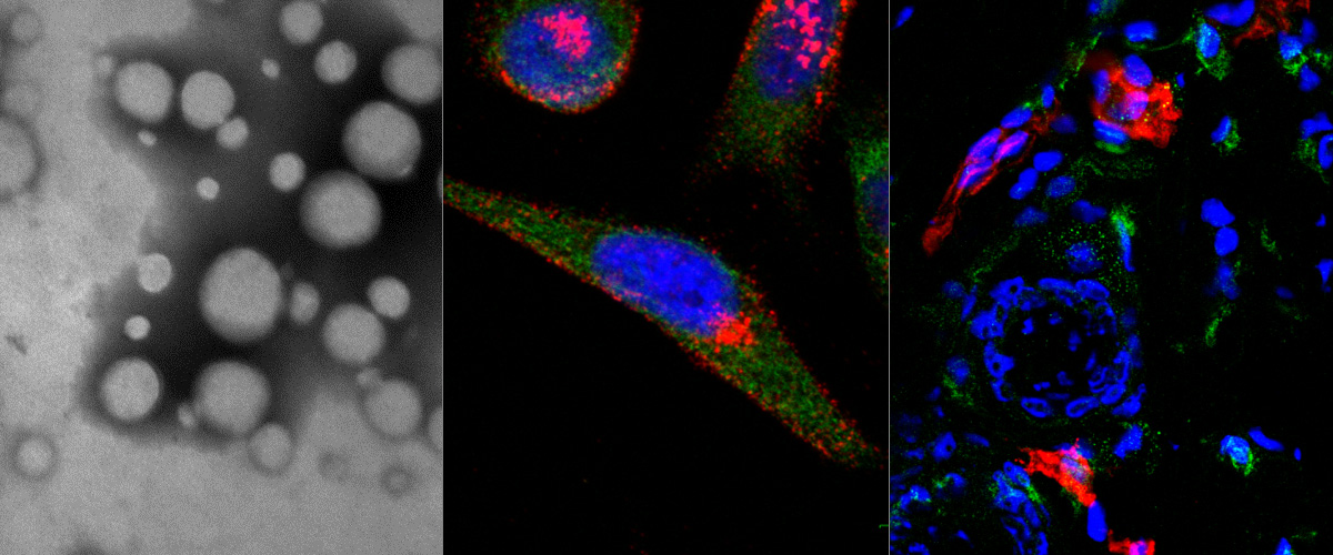
Nanoparticles homing to breast tumor
Left: electron microscopy images of polymeric nanoparticles targeted with tumor penetrating peptide loaded with cancer drug. Right: Confocal microscopy pictures of tumor, kidney, and liver tissue sections of mouse bearing breast tumor i.v. injected with nanoparticles loaded with cancer drug.
Authors: Ines Diaz Bessone PhD, Pablo David Scodeller PhD, Lorena Simon Gracia PhD
Two-photon image of blood vessels with Evans Blue after tail vein injection
Detection with multiphoton microscope Olympus FV1200MPE-BX61WI and Spectra-Physics MaiTai DeepSee IR laser. Objective: XLPLN25xW-MP (NA 1.05; Olympus). Excitation: 950nm. Author: Maarja Haugas, PhD
A peptide for targeted, systemic delivery of imaging and therapeutic compounds into acute brain injuries
Aman P. Mann, Pablo Scodeller, Sazid Hussain, Jinmyoung Joo, Ester Kwon, Gary B. Braun, Tarmo Mölder, Zhi-gang She, Venkata Ramana Kotamraju, Barbara Ranscht, Stan rajewski, Tambet Teesalu, Sangeeta Bhatia, Michael J. Sailor, Erkki Ruoslahti.
Nature Communications 7, Article number: 11980, doi: 10.1038/ncomms11980
Tumor-homing peptides as tools for targeted delivery of payloads to the placenta
King A, Ndifon C, Lui S, Widdows K, Kotamraju VR, Agemy L, Teesalu T, Glazier JD, Cellesi F, Tireli N, Aplin JD, Ruoslahti E, Harris LK.
Science Advances 06 May 2016: Vol. 2, no. 5, e1600349 DOI: 10.1126/sciadv.1600349
Paclitaxel-loaded Polymersomes for Enhanced Intraperitoneal Chemotherapy
Simón-Gracia L,Hunt H, Scodeller PD, Gaitzsch J, Braun GB, Willmore AA, Ruoslahti E, Battaglia G, Teesalu T
Mol Cancer Ther. 2016 Feb 15. pii:molcanther.0713.2015 [Epub ahead of print]
Our ratiometric silver nanoparticles featured on the cover of Nanoscale
Our article titled "Targeted Silver Nanoparticles for Ratiometric Cell Phenotyping" is well received. It has been chosen to the cover of the May issue of Nanoscale and the Atlas of Science has published a layman's summary about the work.
Artwork: Peter and Ryan Allen, UC Santa Barbara, USA.
Urokinase-controlled tumor penetrating peptide
Braun GB, Sugahara KN, Yu OM, Kotamraju VR, Mölder T, Lowy AM, Ruoslahti E, Teesalu T
J Control Release. 2016 Apr 19. pii: S0168-3659(16)30240-1. doi: 10.1016/j.jconrel.2016.04.027. [Epub ahead of print]
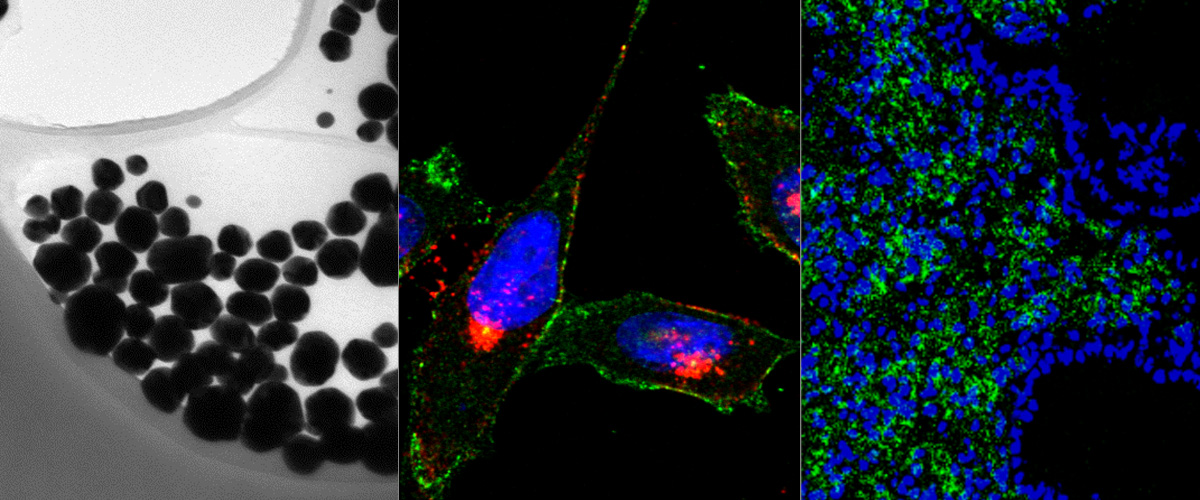
Hollow gold-silver nanospheres
Hollow gold-silver nanospheres imaged with a TEM at 160 000x magnification.
Author: Anne-Mari Anton Willmore, MSc
The "UNO" peptide recognizes circulating tumor associated macrophages
Mouse model of metastatic human gastric cancer, MKN45-P.
Microscope: Olympus FV1200MPE-BX61WI confocal microscope at 20X magnification Green: peptide, Red: tumor associated macrophages, blue: cell nuclei.
Author: Pablo Scodeller, PhD
TT1-FAM-IONW
WT GBM intracranial glioma-bearing mouse was injected with 7,5 mg /kg TT1-FAM-iron oxide nanoworms. After 5 hours of circulation the mouse was perfused and FAM fluorescence was detected by illumatool. Accumulation of particles can be detected in the tumor area.
Author: Pille Säälik, PhD