http://www.ncbi.nlm.nih.gov/pubmed/24907927
Abstract
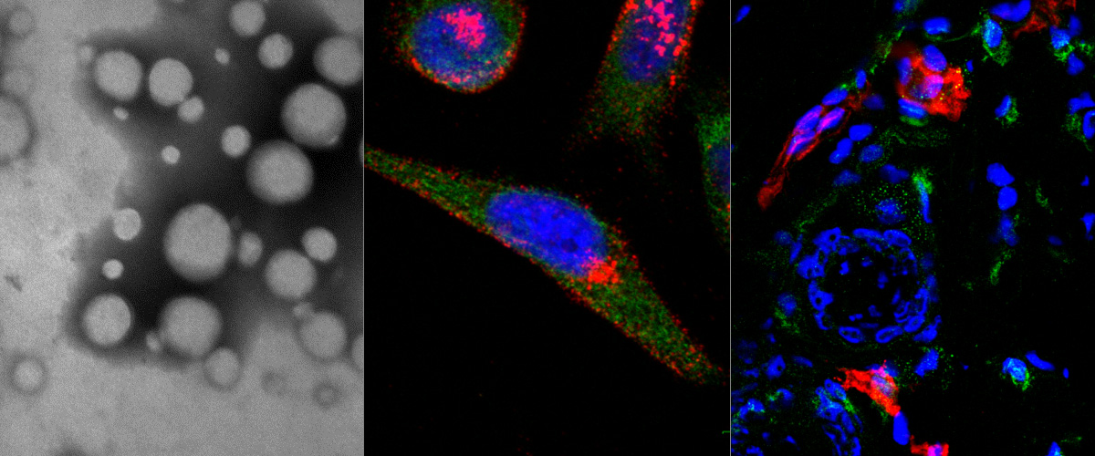
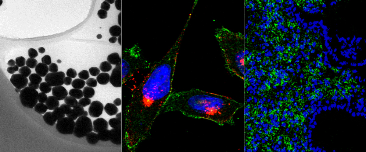
There is considerable interest in using nanoparticles as labels or to deliver drugs and other bioactive compounds to cells in vitro and in vivo. Fluorescent imaging, commonly used to study internalization and subcellular localization of nanoparticles, does not allow unequivocal distinction between cell surface-bound and internalized particles, as there is no methodology to turn particles 'off'. We have developed a simple technique to rapidly remove silver nanoparticles outside living cells, leaving only the internalized pool for imaging or quantification. The silver nanoparticle (AgNP) etching is based on the sensitivity of Ag to a hexacyanoferrate-thiosulphate redox-based destain solution. In demonstration of the technique we present a class of multicoloured plasmonic nanoprobes comprising dye-labelled AgNPs that are exceptionally bright and photostable, carry peptides as model targeting ligands, can be etched rapidly and with minimal toxicity in mice, and that show tumour uptake in vivo.