-
Home

- Pictures
Peptide targeted polymersomes bind to M2 human macrophages
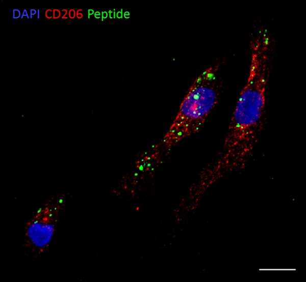
Polymersomes targeted with an M2 macrophage binding peptide (green) identified in our lab using phage display bind to and are taken up by M2s (red) originated from human primary monocytes. Scale bar 10µm. Authors: Anni Lepland, Pablo Scodeller, Lorena Simon Gracia
Polymersomes targeted with an M2 macrophage binding peptide in a mouse model of triple negative breast cancer
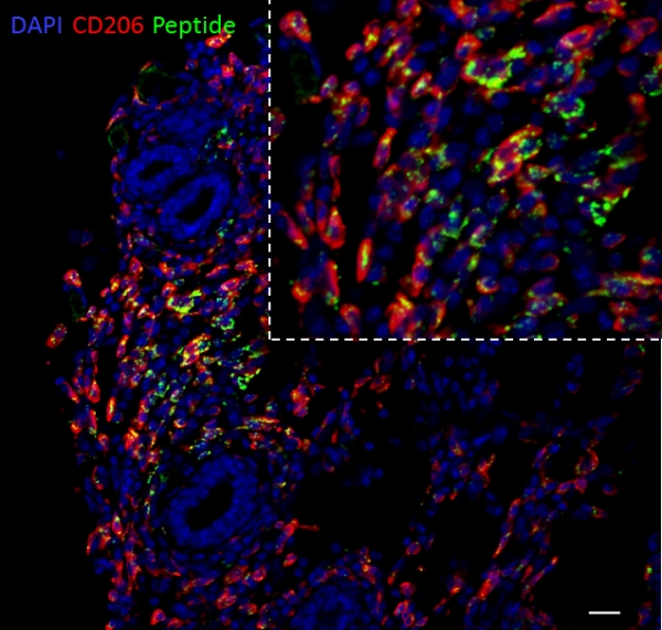
Polymersomes targeted with an M2 macrophage binding peptide (green) identified in our lab using phage display home to M2 TAMs (red) after intravenous administration in a mouse model of triple negative breast cancer. Scale bar 20µm. Authors: Anni Lepland, Pablo Scodeller, Lorena Simon Gracia
High infiltration of CD206+ cells in human breast tumors
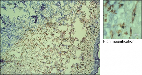
Immunohistochemical staining of human CD206 (brown) with nuclear counterstain (Hematoxilin, in dark blue). Authors: Anni Lepland and Pablo Scodeller.
Long persistence of an M2 TAM internalizing peptide in breast tumors
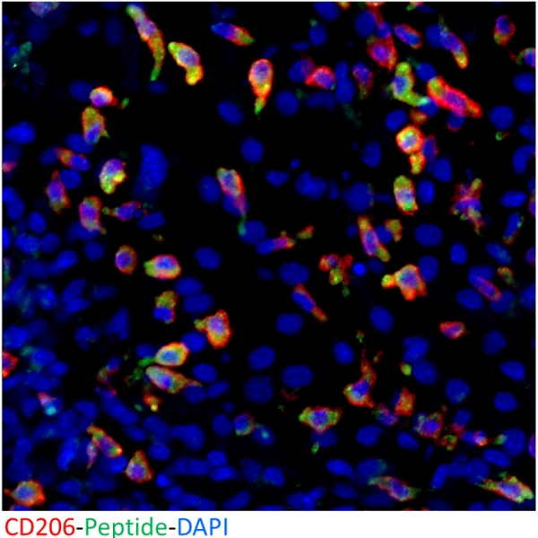
An M2 macrophage binding peptide (green) identified in our lab using phage display remains inside M2 TAMs (red) at least 24 hours after systemic administration in a mouse model of triple negative breast cancer. Authors: Anni Lepland, Pablo Scodeller.
Linear TT1 peptide-coupled FAM-labeled iron oxide nanoparticles home to blood vessels of subcutaneous U87 tumor xenograft
Linear TT1 peptide-coupled FAM-labeled iron oxide nanoparticles (green) home to blood vessels (red) of subcutaneous U87 tumor xenograft. Cell nuclei are stained with DAPI (blue). Scale bar – 50 µm. Author – Pille Säälik PhD
Dark-field image of silver nanoparticles with RPARPAR-MMAE-AgNP internalized by PPC-1 prostate cancer cells
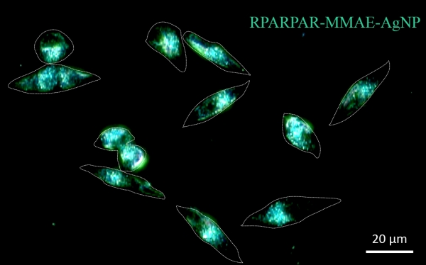
Dark-field image of silver nanoparticles with cytotoxic drug monomethyl auristatin E and RPARPAR peptide (RPARPAR-MMAE-AgNP) internalized by PPC-1 prostate cancer cells after 1 h incubation. Extracellular fraction of AgNPs was removed by etching. Scale bar: 20 μm; dotted lines outline cells. Author: Allan Tobi MSc.
PET-CT image of breast tumor bearing mouse i.v. injected with iRGD targeted nanoparticles
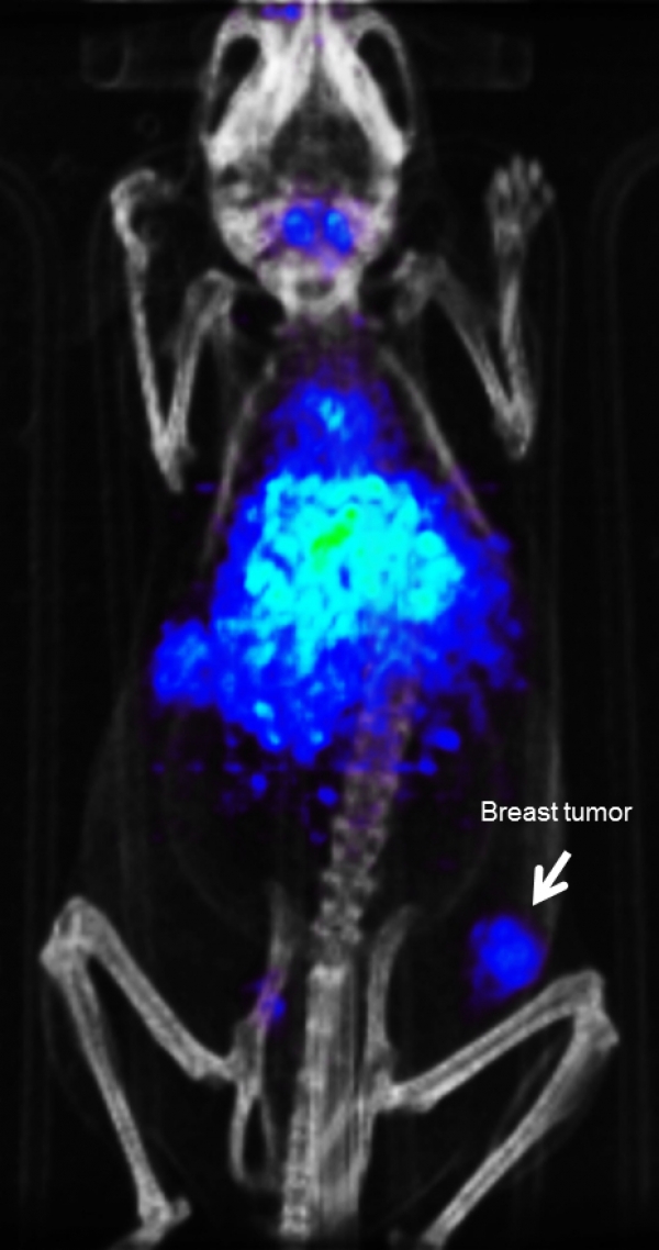
PET-CT scan was acquired 24 hours after sample administration. The radiolabeled iRGD-nanoparticles accumulates in breast tumor (white arrow). Authors: Lorena Simón Gracia and Pablo Scodeller
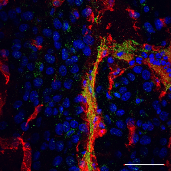
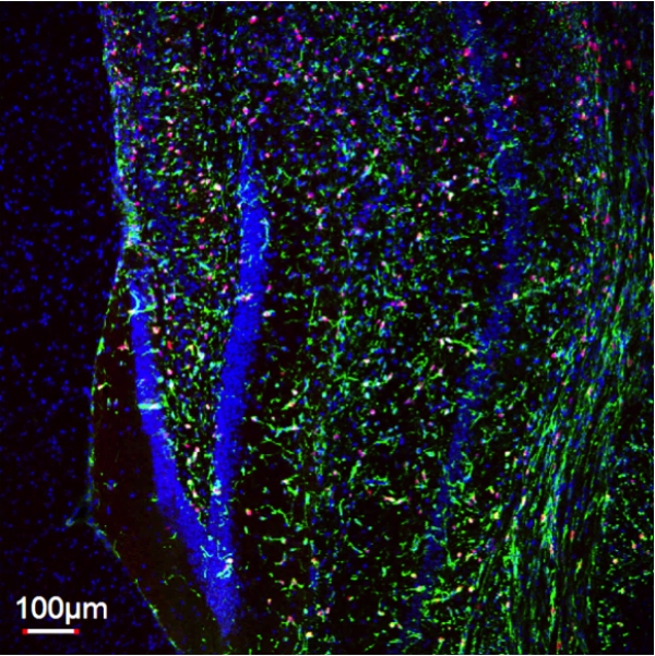
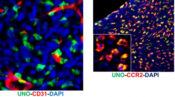
FAM-UNO homes to TIE2+ tumor macrophages
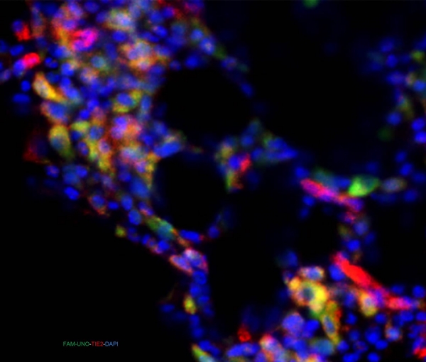
Mouse model of triple negative breast cancer, 4T1. Microscope: Olympus FV1200MPE-BX61WI confocal microscope at 20X magnification Green: peptide, Red: TIE2, blue: cell nuclei. Intravenous injection. Author: Pablo Scodeller
Circular structure of blood vessels homing FAM-labeled nanoparticles in P3-stem like glioblastoma
Circular structure of blood vessels homing FAM-labeled nanoparticles in P3-stem like glioblastoma. CD31 (red), FAM NPs (green), nuclei (blue). Author: Maarja Haugas, PhD
Infiltrating CD206-positive macrophages in peritoneal MKN45P gastric carcinoma xenograft
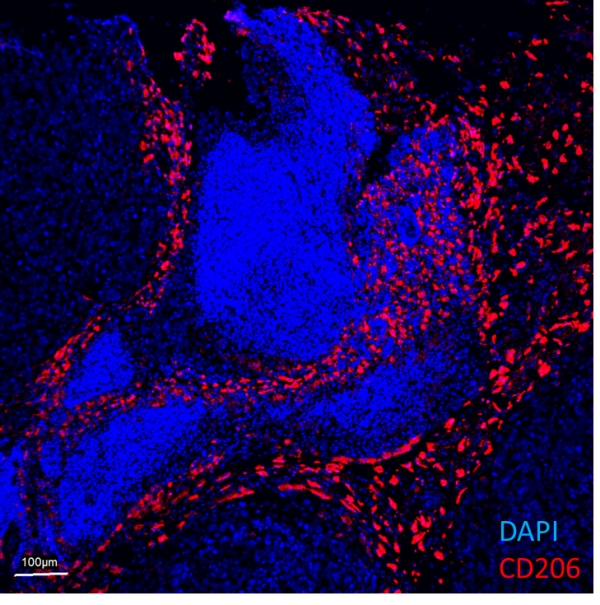
Infiltrating CD206-positive macrophages in peritoneal MKN45P gastric carcinoma xenograft.
Author: Hedi Hunt, MSc.
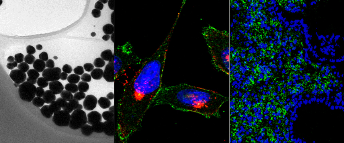
Transmission electron microscopy image of a dextran coated iron oxide nanoworm structure
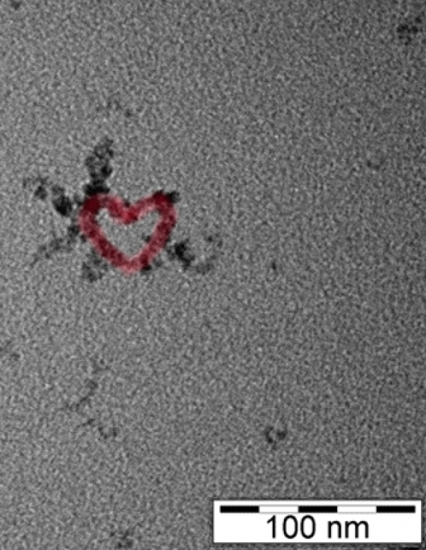
Red color was applied with photo editing. Merry Christmas!
Author: Allan Tobi, MSc
Glih27 peptide conjugated with iron oxide nanoworms (green) in mouse brain with VEGF KO glioblastoma
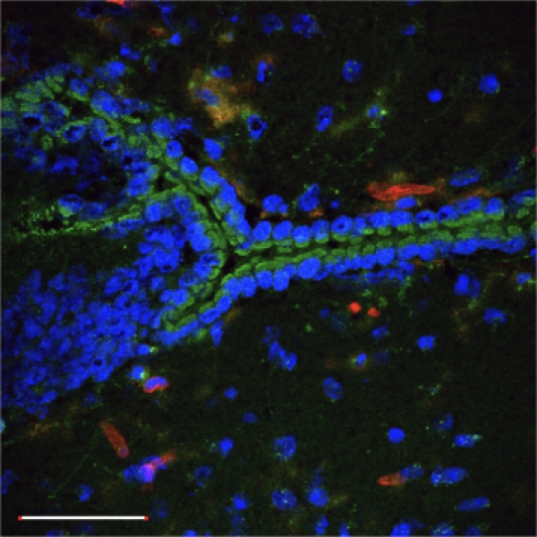
The nanoworms with peptide home to ependymal cells. Red – blood vessels, blue – DAPI.
Author: Pille Säälik, PhD
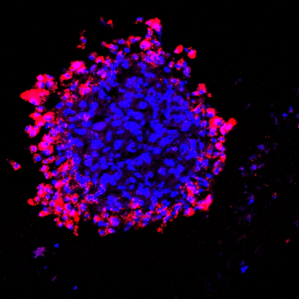
Targeting-polymeric nanoparticles are internalized in MCF-7 and U87 spheroids
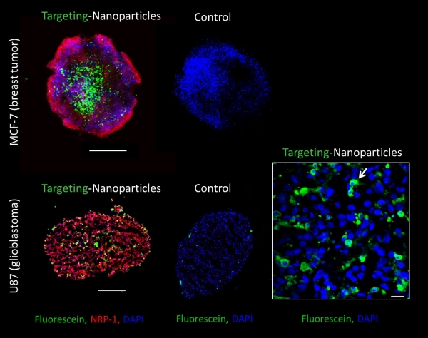
Fluorescein-labeled polymeric nanoparticles functionalized with a targeting peptide (in green) are internalized into MCF-7 (from breast tumor) and U87 (from glioblastoma) spheroids that express the NRP-1 receptor (in red). The nanoparticles are observed inside the U87 cells (white arrow). Scale bar are 200 µm and 20 µm (in the big magnification).
Authors: Lorena Simón-Gracia, Ines Diaz Bessone, Kadri Toome
An infiltratively growing patient-derived mouse glioma xenograft P21 developed in our lab expresses nestin (green) and Ki67 (red) positive cells.
Cell nuclei are stained with DAPI (blue).
Author: Pille Säälik, PhD
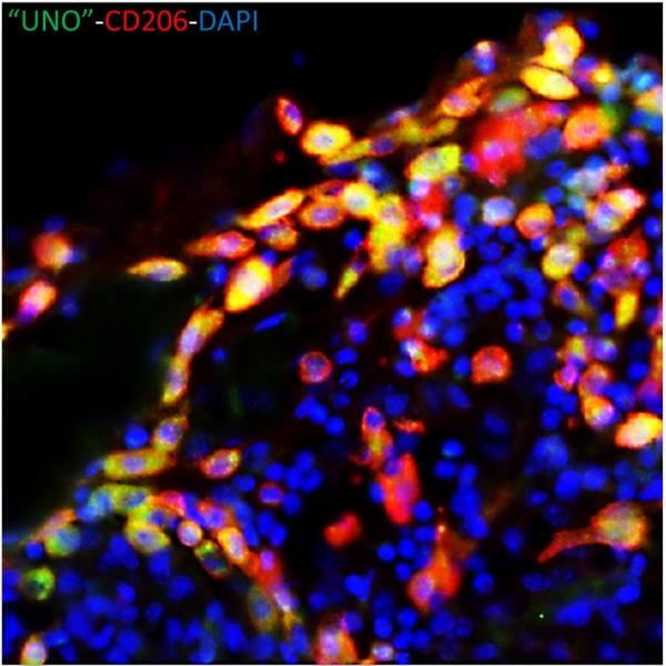
Intravenously injected "UNO" peptide enters protumoral macrophages
Angiogenic glioblastoma, WTGBM.
Microscope: Olympus FV1200MPE-BX61WI confocal microscope at 20X magnification Green: peptide, Red: tumor associated macrophages, blue: cell nuclei.
Authors: Pablo Scodeller PhD, Pille Säälik PhD
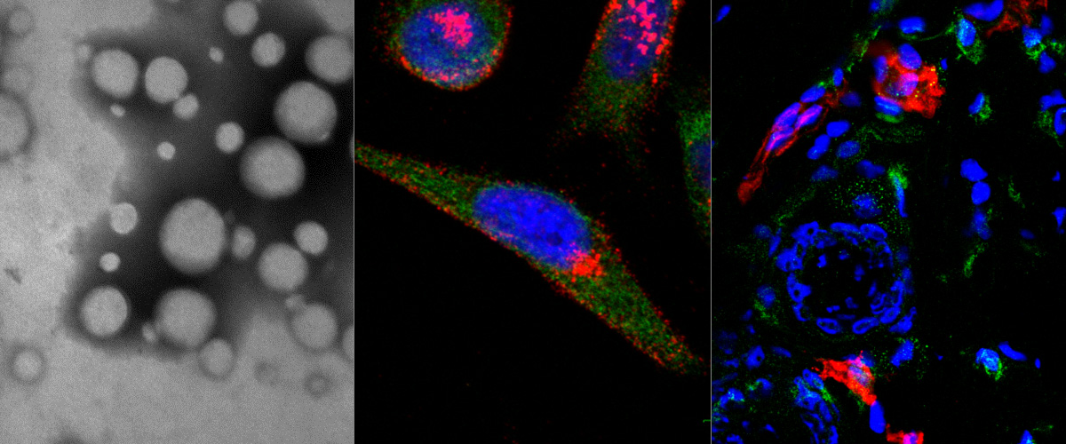
Nanoparticles homing to breast tumor
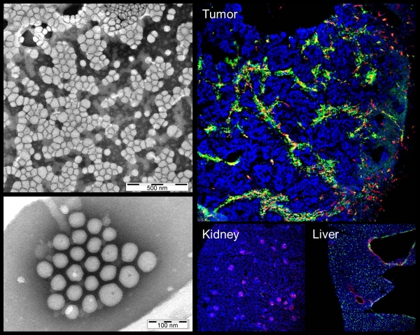
Left: electron microscopy images of polymeric nanoparticles targeted with tumor penetrating peptide loaded with cancer drug. Right: Confocal microscopy pictures of tumor, kidney, and liver tissue sections of mouse bearing breast tumor i.v. injected with nanoparticles loaded with cancer drug.
Authors: Ines Diaz Bessone PhD, Pablo David Scodeller PhD, Lorena Simon Gracia PhD
Two-photon image of blood vessels with Evans Blue after tail vein injection
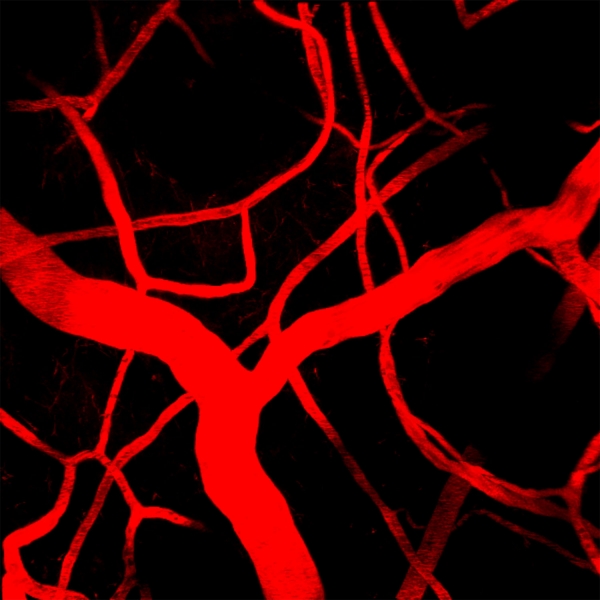
Detection with multiphoton microscope Olympus FV1200MPE-BX61WI and Spectra-Physics MaiTai DeepSee IR laser. Objective: XLPLN25xW-MP (NA 1.05; Olympus). Excitation: 950nm. Author: Maarja Haugas, PhD
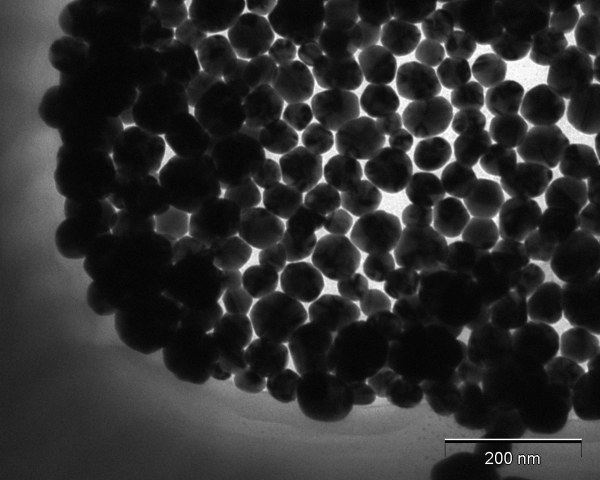
Hollow gold-silver nanospheres
Hollow gold-silver nanospheres imaged with a TEM at 160 000x magnification.
Author: Anne-Mari Anton Willmore, MSc
The "UNO" peptide recognizes circulating tumor associated macrophages
Mouse model of metastatic human gastric cancer, MKN45-P.
Microscope: Olympus FV1200MPE-BX61WI confocal microscope at 20X magnification Green: peptide, Red: tumor associated macrophages, blue: cell nuclei.
Author: Pablo Scodeller, PhD
TT1-FAM-IONW
WT GBM intracranial glioma-bearing mouse was injected with 7,5 mg /kg TT1-FAM-iron oxide nanoworms. After 5 hours of circulation the mouse was perfused and FAM fluorescence was detected by illumatool. Accumulation of particles can be detected in the tumor area.
Author: Pille Säälik, PhD
A peptide identified in the lab using in vivo phage display homes to tumor macrophages in a mouse model of a highly metastatic human gastric cancer, MKN45-P
Microscope: Olympus FV1200MPE-BX61WI confocal microscope at 60X magnification
Green: peptide, Red: macrophages, blue: cell nuclei.
Author: Pablo David Scodeller, PhD
IONW cycle of life
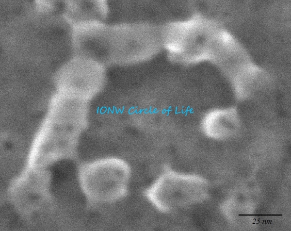
Microscope: SEM (magnification: 600,000x).
Author: Allan Tobi, MSc student
Expression of stem cell markers Aldh1a1 and Nestin in human stem cell like glioblastoma cell line P3
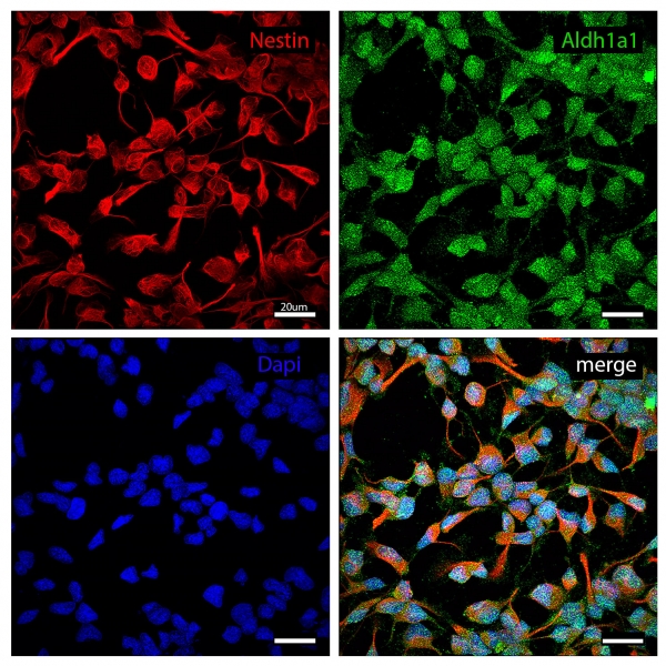
Confocal detection with Olympus FV1200MPE-BX61WI microscope and 60xoil objective.
Author: Maarja Haugas, PhD
Ex vivo visualization of targeted nanoworms homing to intraperitoneal and subcutaneous tumors after i.v. injection
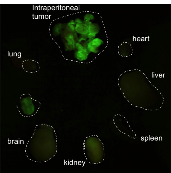
Mouse model: peritoneal gastric carcinoma ( MKN-45 in Nude mice).
Author: Hedi Hunt, MSc
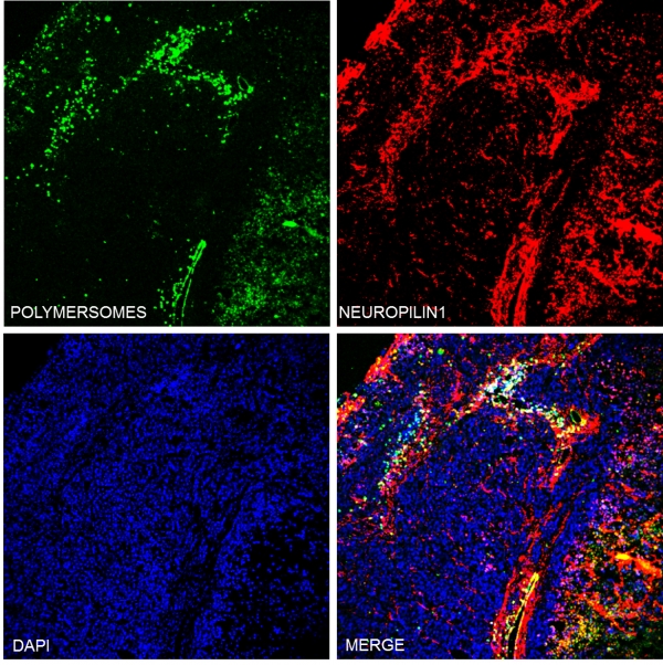
Neuropilin 1 expression (in red) in glioblastoma cell lines used in our lab
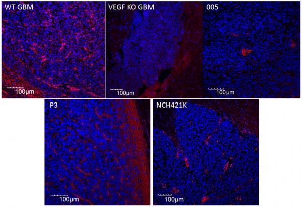
WT GBM, VEGF KO GBM and 005 – mouse-derived; P3 and NCH421K – human-derived cell lines. Blue – nuclei (DAPI).
Image acqusition: confocal system Olympus FV1200MPE.
Author: Pille Säälik, PhD
Second harmonic generation image of collagen fibers in mouse dermis
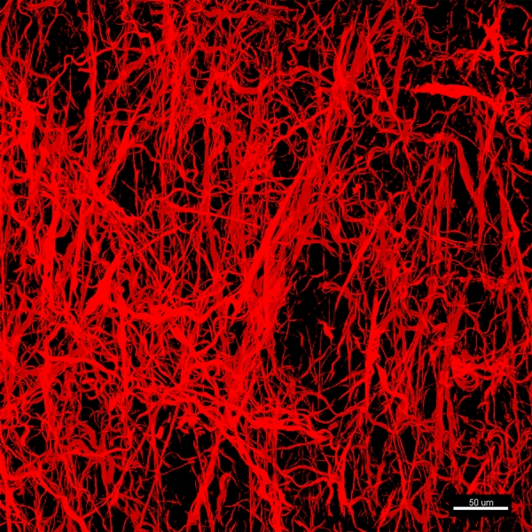
Detection in wavelength of 800nm with Spectra Physics MaiTai Deep See Ti:Sapphire laser. Multiphoton microscope Olympus FV1200MPE; objective XLPLN 25xW-MP (NA 1.05).
Author: Maarja Haugas, PhD
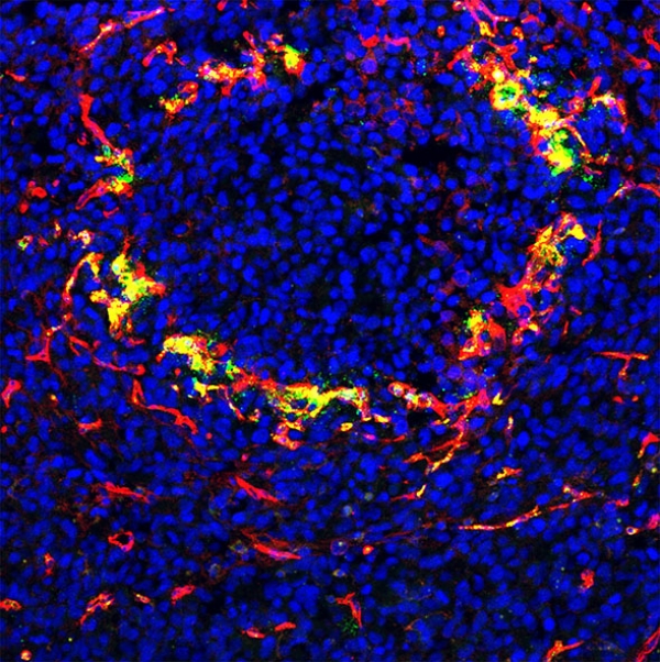
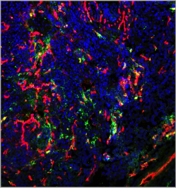
Targeted polymersomes penetrate deep into peritoneal tumors
Targeted polymersomes (in green) target Neuropilin1 rich regions (in red) in gastric peritoneal tumors when injected intraperitoneally.
Authors: Lorena Simon Gracia, PhD and Pablo Scodeller, PhD
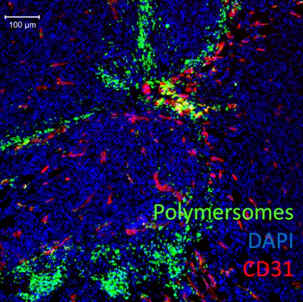
Homing of nanoworm nanoparticles to peritoneal gastric carcinoma ( MKN-45 Nude mice)
Sample collected 5h after i.p. injection.
Red: Vessels
Green: NW-TT1-FAM
Blue: Nuclei
Technology: Confocal Microscopy (Olympus FV1200MPE)
Author: Hedi Hunt, MSc
Homing of polymersome nanoparticles to peritoneal colon carcinoma (CT26 Balb/c syngeneic tumor model, sample collected 24 hours after injection)
Technology: confocal microscopy
Authors: Lorena Simon Gracia, PhD and Hedi Hunt, MSc
Ali Baba and fourty thieves a.k.a 41 T7 bacteriophages amplifying on the E. coli agar plate.
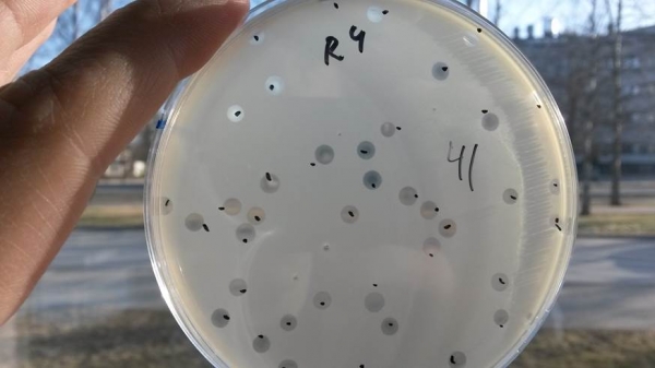
Author: Tarmo Mölder, MSc
U87 cell line spheroids taking up RPARPAR-AgNPs (red)
Technology: Confocal microscopy 20x
Author: Kadri Toome, MSc
In vivo imaging of a mouse bearing luciferase-expressing MKN-45 gastric carcinoma.
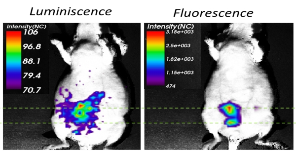
Left: luminiscence of the gastric tumor. Right: fluorescence of polymeric vesicles homing to the tumor.
Images were acquired using ART OPTIX MX3 imaging system.
Authors: Lorena Simón Gracia, PhD and Pablo Scodeller, PhD
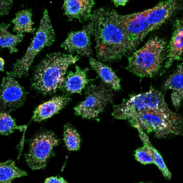
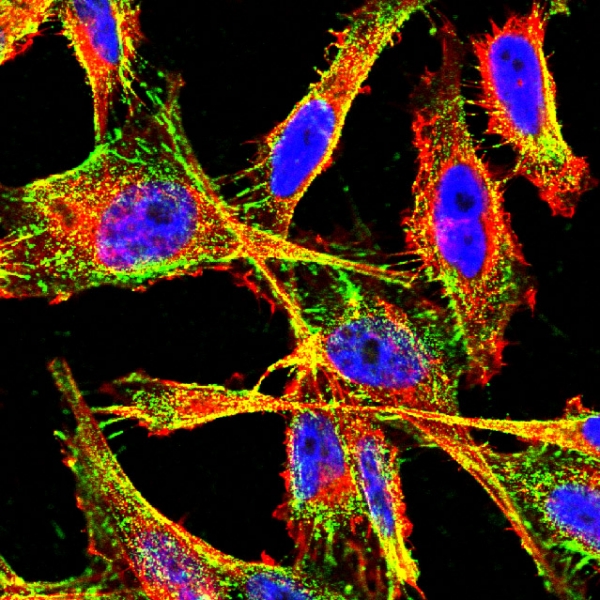
SKOV3 ovarian cancer cells
Technical information: PS-Rho-iRGD-FAM
Polymersomes-Red
iRGD peptide -Green
NRP-1-Purple
Nuclei-Blue
Author: Hedi Hunt, MSc
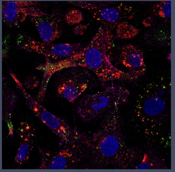
Co-culture of NRP1 (green) expressing PPC1 cells along with M21 cells; both cell lines express p32 (white), RPARPAR-AgNPs (red) are taken up by PPC1 cells.
Technical information: Confocal microscopy 63x.
Author: Anne-Mari Anton Willmore, Lorena Simon Gracia
PC3, prostate cancer cells, taking up RPARPAR-AgNPs (red), also showing NRP1 expression (green) and p32 expression (white)
Technical information: Confocal microscopy, 63x magnification.
Author: Anne-Mari Anton Willmore, MSc
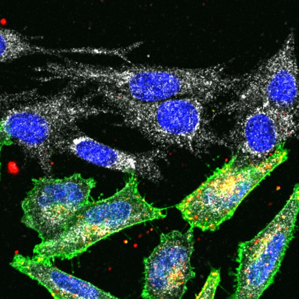
Selective internalization of RPARPAR silver nanoparticles (Ag-RPARPAR) in NRP-1 expressing cells. In mixed culture, Ag-RPARPAR are taken by NRP-1 (green)-expressing PPC1 prostate carcinoma cells but not by M21 melanoma cells not expressing NRP-1.
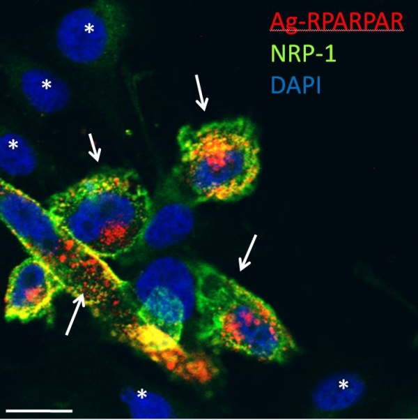
Technical information: Confocal microscopy, 63x magnification.
Authors: Lorena Simon Gracia, PhD and Anne-Mari Anton Willmore, MSc
Confocal microscopy picture showing the internalization of polymersomes labeled with Rhodamine and targeted with RPARPAR-FAM peptide in PPC-1 cells. 63 x magnification.
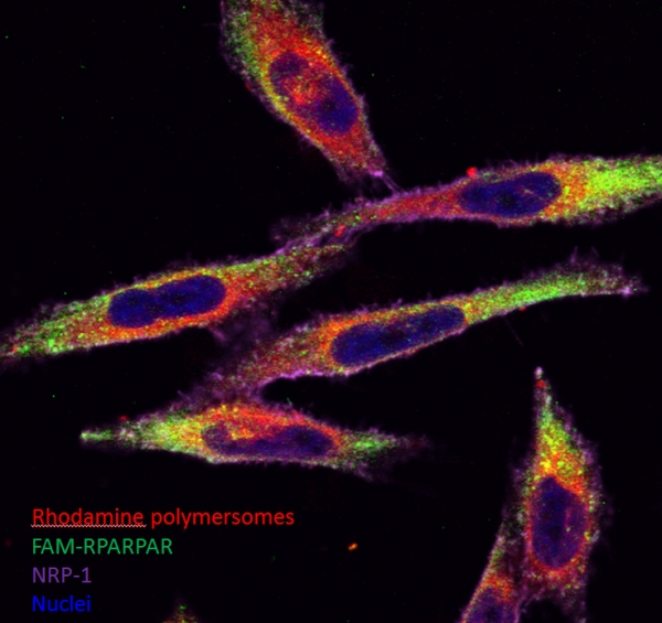
Authors: Lorena Simon Garcia, PhD and Hedi Hunt, MSc
Transmission electron microscope image of hybrid silver/polymeric nanoparticles (NPs)
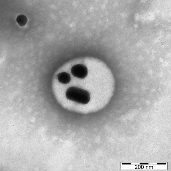
Ag-PVP NPs are encapsulated inside POEGMA-PDPA polymersomes. pH-sensitive polymersomes release the AgNPs and encapsulated drug inside the tumor cells. Targeted hybrid NPs can be used as a theranostic tool.
Author: Lorena Simón Gracia, PhD
Penetration of uCendR-RGD peptide to 4T1 breast tumors in vivo
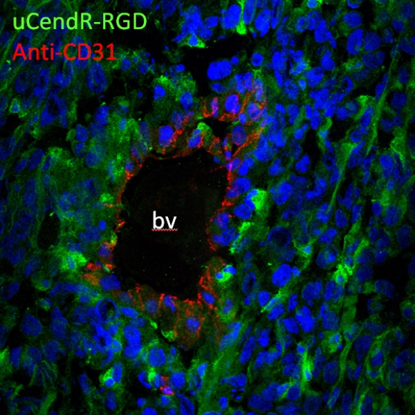
uCendR peptide was designed by positioning the cryptic CendR motif in the context of recognition and cleavage site of a tumor-associated protease, urokinase-type plasminogen activator (uPA). FAM-labeled uCendR peptide containing additional RGD vascular recruitment module was injected into 4T1breast tumor mice, after 3hr circulation the mice were perfused with PBS and tumors sectioned for microscopy. Confocal microscopy confirmed the accumulation of synthetic uCendR-RGD peptide (green) in tumor tissue, and revealed extravasation and spreading within the tumor. CD31- positive tumor blood vessels (bv) – red.
Author: Tambet Teesalu, PhD
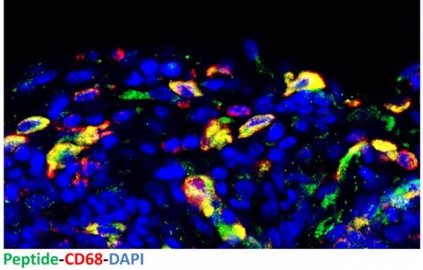
Intravenously injected phage displaying a candidate glioma homing peptide (green) colocalizes with CD68-positive glioma macrophages (red), nuclei-blue.
Model: VEGF KO GBM, collaboration with Gabriele Bergers (UCSF).
Authors: Kadri Toome, MSc and Pille Säälik, PhD
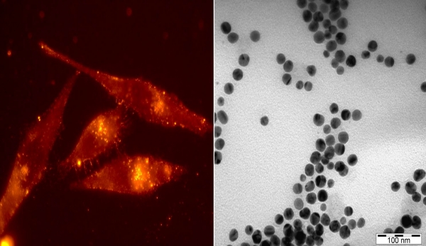
109Ag nanoparticles as seen with TEM 210k X and coupled with fluorescent dye and CendR peptide being taken up by cancer cells 100 X
Author: Anne-Mari Willmore, MSc
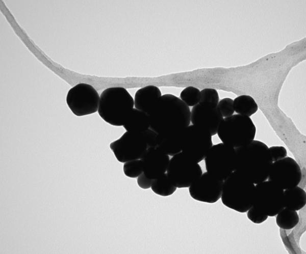
TEM picture of 20 nm silver nanoparticles at 65,000x of magnification
Author: Lorena Simon Gracia, PhD
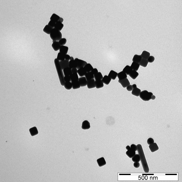
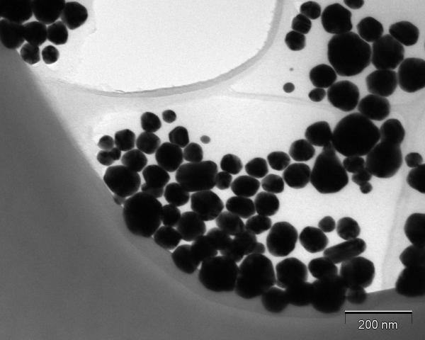
TEM image of silver, palladium nanoparticles at 93,000x magnification
Author: Anne-Mari Willmore, MSc
Internalization of prototypic CendR (RPARPAR) phage by PPC1 prostate carcinoma cells in culture
Phage – green, phalloidin – red, DAPI-blue
Aithor: Tambet Teesalu, PhD
TEM image of AgPVP nanoparticles at 73,000x magnification
Author: Lorena Simón Gracia, PhD
TEM image of PEG-PDPA Polymersomes at 105,000x magnification
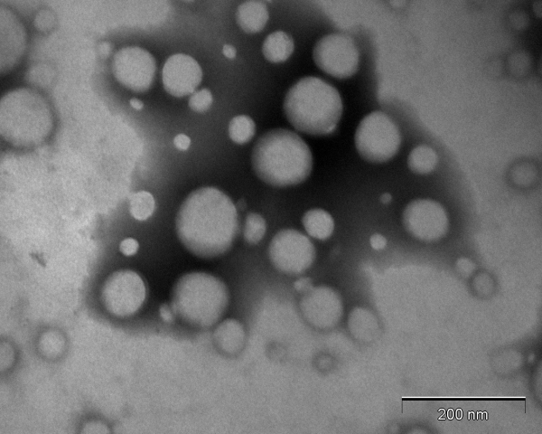
Author: Lorena Simón Gracia, PhD
AgPVP nanoparticles at 105,000x magnification
Author: Anne-Mari Willmore, MSc